Why imaging in the near-infrared?
Imaging of thick sample such as a piece of tissue requires efficient light collection. However, the tissue penetration depth is a major problem that prevents us from using a conventional confocal microscope to image arbitrarily specimen. Both absorption and scattering of light, which are wavelength-dependent, will prevent light from being a tight focus where can detect the emitted light. Thus, imaging in the near-infrared:
-minimize absorption
-reduce the scattering.
One possibility to image in the infrared is using infrared dyes. The other approach is to use two-photon microscopy and image standard dyes in the infrared.
Two-photon microscopy
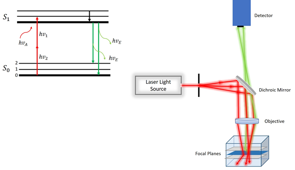
Two photons microscopy is another family of optical sectioning. In this technique, two photons are of half energy to excite dye at almost the same time. By adding up those two half-energy photons we can achieve full excitation (Figure 1). Multiphoton microscopy allows characterizing the samples (both biological and non-biological) of micrometre size with better resolution and minimum or no photodegradation.
This process depends on the square of the intensity of the light hitting sample. It means that out-of-focus light is much dimmer than in-focus light is not going to excite any species to an appreciable level. In other words, only the excited spot is at the focal spot of confocal. Two photons microscope does not require pinhole because we only get light from in-focus spot there is no out-of-focus light. Due to efficient light collection, this technique works well for thicker samples such as a piece of tissue.
The following picture shows the principle of two photon microscopy.

Setup:
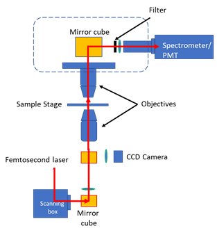
the samples kept on the sample stage are usually excited by ultrashort laser pulses to show multiphoton fluorescence induced by higher-order optical nonlinearities of the sample. This fluorescence signal containing rich information about the sample can be fed into a spectrometer for spectral decomposition. The scanning box provided along with the automatic controller allows the raster scanning of samples to obtain the images of active multiphoton regions in either transmission or reflection mode.
Image by roster scanning:
for creating the image we use a laser which collimated and can be focused to a tight spot on the sample. By scanning laser point by point over sample and recording intensity at each spot we can reconstruct an image from all these individual spots. By changing the entrance angle of illumination, we can illuminate a different spot on the sample. One scanning mirror is used to scan different spots on the sample as well as bringing emission light that comes back from the sample. Mirror change the angle at which laser hits the sample and accordingly change the position in the sample we are imaging.
The microscope is equipped with a Halogen lamp, CCD camera, two PMTs, objective lenses and filter turrets.
