Introduction
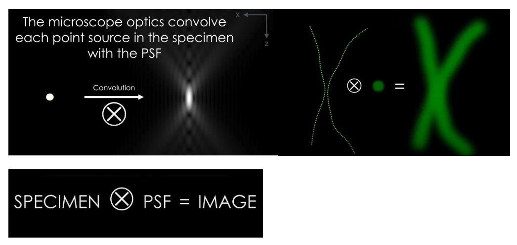
The point spread function (PSF) is the three-dimensional image of a point-like object under the microscope. It describes how a microscope blurs out any point-like object due to the diffraction of light, which determines the microscope's resolution limit. The PSF can be used to evaluate the axial resolution and the lateral resolution of a microscope. The axial resolution is the minimum distance that two points along the optical axis can approach each other and still be resolved, while the lateral resolution is the minimum distance that two points in the same focal plane can approach each other and still be resolved. The PSF is influenced by several factors, such as the numerical aperture of the objective lens and the condenser, the wavelength of light used, the refractive index of the medium, and the alignment and quality of the optical components. Higher numerical aperture, shorter wavelength, higher refractive index, and better alignment and quality can improve the PSF and thus increase the resolution of a microscope.
Microscopy resolution is the ability of a microscope to distinguish details of a specimen or sample. It can also be defined as the smallest distance between two points on a specimen that can still be distinguished as separate entities. The smaller this value, the higher the resolving power of the microscope and the better the clarity and detail of the image. The theoretical limit of resolution for a light microscope is about 200 nm, which is much larger than the size of many molecules and atoms. This physical limit is known as the Abbe limit, named after Ernst Abbe, who first proposed this concept in 1873.
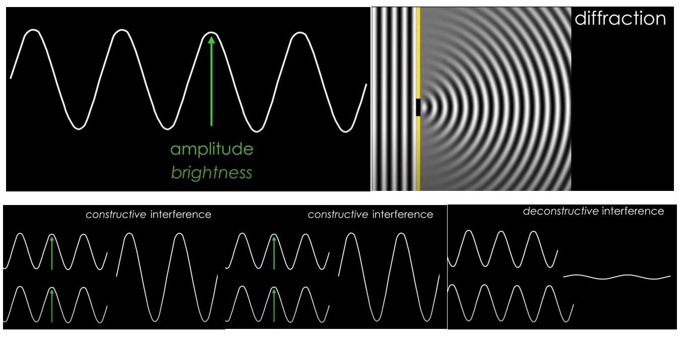
One factor that limits the resolution of light microscopy is diffraction, which is the bending of light as it passes through a small opening or around an obstacle. Diffraction results in what we call the PSF. Diffraction causes light to spread out and form interference patterns, which blur the image of the specimen. Diffraction is more noticeable when the aperture (the opening that controls how much light enters the lens) is small. The diffraction pattern for a circular aperture has a central bright spot surrounded by concentric rings of light, called an Airy disc. The size of the Airy disc depends on the wavelength of light and the numerical aperture (a measure of how much light a lens can collect) of the lens.
Close look at a PSF
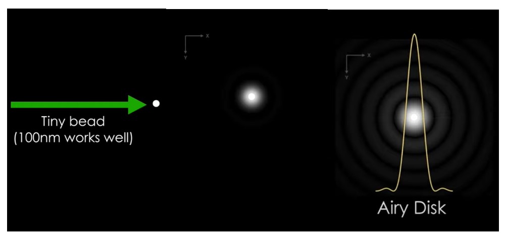
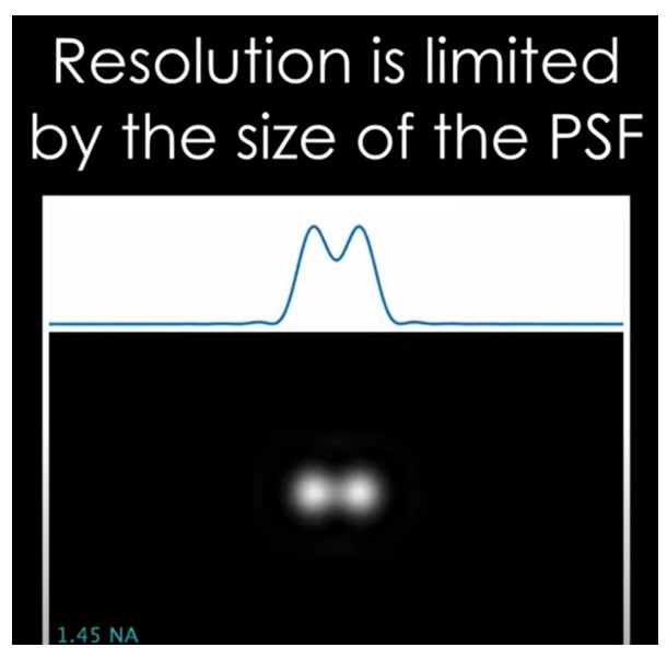
When a microscope is used to form an image of a tiny point source of light. The point spread function defines what the image of that point source will look like. To get the most accurate view of the point spread function, you need to look at a single point source of light that is below the theoretical resolution limit of the microscope. Let us imagine we are imaging a fluorescent bead a sphere that’s in fused with fluorophore and that is below the resolution limit of the microscope we actually use fluorescent beads when we want to empirically measure the PSF of a microscope. When we look at the lateral optical image of the bead, we are going to see a spot that is larger than the bead itself and it has a blurry edge. If we enhance the image contrast, we can see weaker intensity values, and the bright centre is surrounded dark and light concentric rings. If we draw a line across this image and look at the intensity of the image across that line, we will see that the bright center which we call the maxima has a gaussian-like distribution and that the outer rings are much dimmer that maxima.
PSF results from diffraction & interference
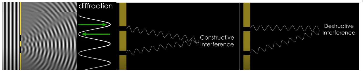
Diffraction is the bending of waves when they pass through a narrow aperture or around an obstacle. It occurs because waves can spread out and interfere with each other after passing through the aperture or obstacle. Diffraction is a wave characteristic that occurs for all types of waves, such as laser, sound, water, and matter waves. Interference is the addition of two or more waves that result in a new wave pattern. It occurs when waves overlap and combine constructively or destructively depending on their phase difference. Interference is also a wave characteristic that occurs for all types of waves. The difference between diffraction and interference is that diffraction involves the bending of a single wave due to an aperture or obstacle, while interference involves the combination of two or more waves due to their superposition. Diffraction and interference often occur together, such as in the case of a diffraction grating or a double-slit experiment.
Diffraction in the microscope
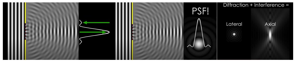
Diffraction in the microscope is a phenomenon that occurs when light waves pass near a barrier, such as an aperture or a specimen, and bend around it. Diffraction causes the light waves to spread out and interfere with each other, forming a diffraction pattern. Diffraction limits the resolution of the microscope, because it prevents the image of a point source or object from being infinitely sharp. Instead, the image of a point source or object is blurred into a finite area called the PSF. The degree of diffraction depends on the wavelength of light and the numerical aperture (NA) of the objective lens. Shorter wavelengths and higher NA values result in less diffraction and better resolution. The resolution limit of a microscope is approximately half the wavelength of light divided by the NA. This limit is also known as the diffraction barrier. To overcome this barrier, some super-resolution microscopy techniques use special methods to manipulate the diffraction pattern or to localize single molecules beyond the diffraction limit.
The effect of the PSF on your images
The effect of the PSF on your images is that it reduces the sharpness and contrast of the details in your specimen. The PSF causes the image of a point source or object to be blurred into a finite area that is larger than the actual size of the source or object. This means that if two-point sources or objects are closer than the size of the PSF, they cannot be resolved as separate entities in the image. The PSF also affects the intensity or brightness of the image, because it spreads out the light from a point source or object over a large area. The PSF can also introduce artifacts such as ringing or halo effects around sharp edges or bright features in the image.
Resolution equations
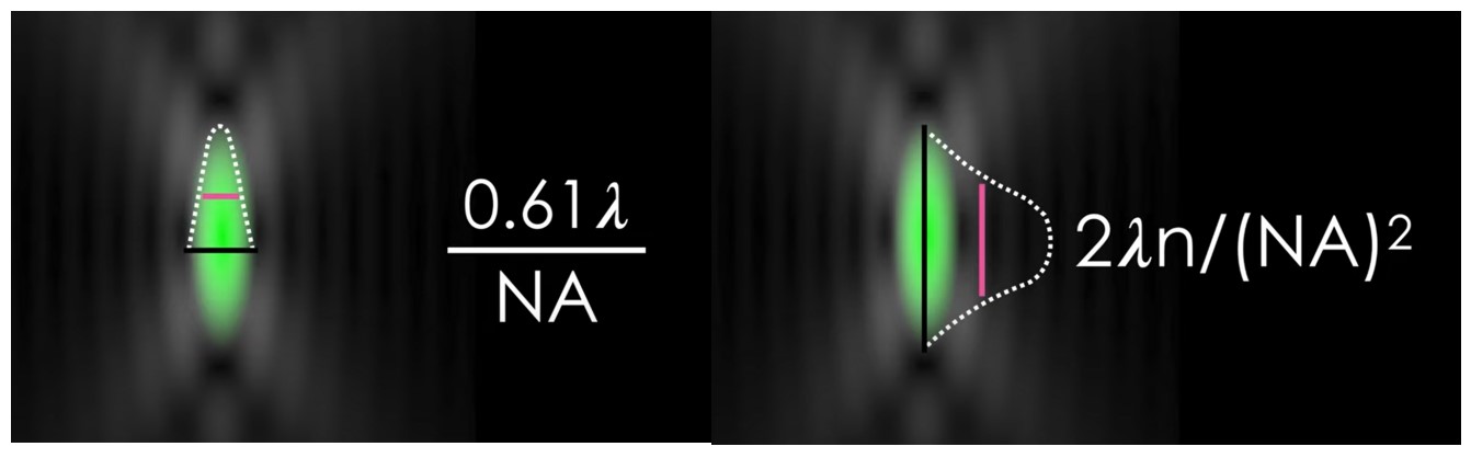
There are different formulas for calculating the resolution of a microscope, depending on the assumptions and factors involved. One of the most common formulas is the Ernst Abbe formula, which is given by:
• Resolution (r) = 0.61λ/NA
Where r is the resolution, λ is the wavelength of light coming from the object, and NA is the numerical aperture of the objective lens. This formula assumes that the numerical aperture of the condenser lens is equal to or greater than that of the objective lens, and that the light source is coherent and monochromatic. Another formula that takes into account the numerical aperture of both the objective and condenser lenses is:
• Resolution (r) = 1.22λ/ (NA (obj) + NA (cond))
Where r is the resolution, λ is the wavelength of light coming from the object, NA (obj) is the numerical aperture of the objective lens, and NA (cond) is the numerical aperture of the condenser lens. This formula assumes that the light source is incoherent and polychromatic. Both formulas show that resolution is inversely proportional to wavelength and directly proportional to numerical aperture. Therefore, to improve resolution, one should use shorter wavelengths and higher numerical apertures.
Rayleigh criterion
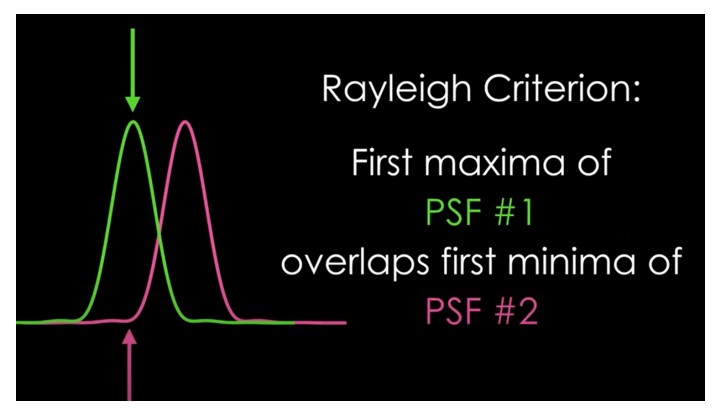
The Rayleigh criterion is a rule that defines the minimum angular separation between two point sources or objects that can be resolved by an imaging system. The criterion is based on the diffraction pattern produced by a circular aperture, which has a central maximum surrounded by smaller maxima. According to the Rayleigh criterion, two-point sources or objects are just resolved when the central maximum of one source coincides with the first minimum of the other source.
The angular separation that satisfies this condition is given by the formula:
• θ = 1.22λ /D
where θ is the angular separation, λ is the wavelength of light, and D is the diameter of the aperture. The Rayleigh criterion can be used to determine the resolution limit of optical instruments such as microscopes, telescopes, and cameras. The smaller the value of θ, the better the resolution. The resolution can be improved by using shorter wavelengths of light or larger apertures.
Size of PSF
The size of the PSF depends on several factors, such as the wavelength of light, the numerical aperture of the imaging system, and the shape and size of the aperture. Generally, the PSF is smaller for shorter wavelengths of light and larger for longer wavelengths of light. The PSF is also smaller for higher numerical apertures and larger for lower numerical apertures. The PSF can also have different shapes depending on the shape of the aperture. For example, a circular aperture produces an Airy disk pattern, while a rectangular aperture produces a sinc function pattern. The size of the PSF can be measured by imaging a point source or object that is smaller than the resolution limit of the imaging system, such as a sub-wavelength bead or a single molecule. The diameter or width of the central maximum of the PSF can be used as an estimate of the resolution limit. Alternatively, the size of the PSF can be calculated using formulas based on diffraction theory or optical transfer function.
Convolution
Image convolution is a process of applying a filter or a kernel to an image in order to modify or enhance the image. A filter or a kernel is a small matrix that contains some values that determine how the image is transformed. Image convolution involves multiplying each element of the filter with the corresponding element of the image and then adding them up to get a new value for the output image. This operation is repeated for every pixel in the image. Image convolution can be used for various purposes, such as blurring, sharpening, edge detection, noise reduction, and more. Different filters or kernels can produce different effects on the image. For example, an identity filter does not change the image, a box blur filter averages the neighboring pixels to create a smoothing effect, and a Laplacian filter highlights the edges or boundaries in the image.
Disclaimer: The pictures come from the Internet, if there is any infringement, please contact to delete the post.