LSCM(Laser Scanning Confocal Microscopy) is a high-tech optical and electronic instrument developed from the 1980s. In recent years, the technology has developed rapidly and its functions have been improved day by day. It is one of the most advanced fluorescence imaging and cell analysis means in the field of biomedicine and chemistry, and its application fields are becoming more and more extensive. It takes laser as the light source, and is composed of confocal imaging scanning system, electronic-optical system and microcomputer image analysis system. The light beam after focusing falls on the sample (tissue thick sheet or cell) at different depths of the tiny point, and mobile scanning, through the color development of electrical signals, can make the sample any point of the reflected light of the image, are accurately received and generated signals, transferred to the color display, and then connected to the computer image analysis system for analysis and processing.
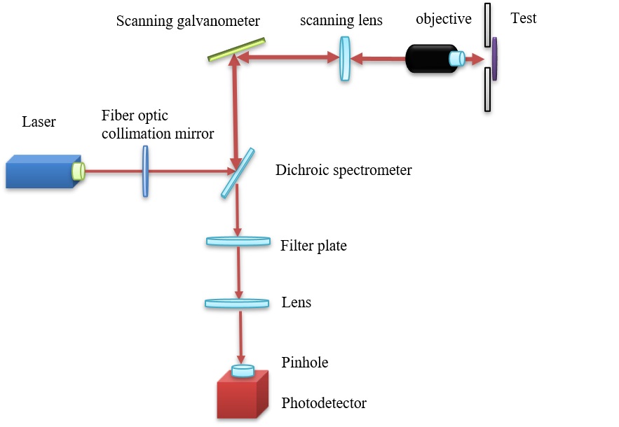
The light source of LSCM system is ion laser or helium-neon laser. The laser beam from the light source passes through the lighting pinhole and becomes point light source P. The point light source P enters the imaging system of the high numerical aperture microscope objective through the beam splitter and forms the image point P in the partially transparent sample under test. The scattered light, reflected light and other fluorescence emitted from the image point P pass through the objective lens and spectroscope in the opposite direction and then incident on the single point detector. The electrical signal received by the detector is finally processed by a computer.
|
| Laser scanning confocal imaging schematic |
Confocal microscope is a kind of special fluorescence microscope with three conjugated points compared to the ordinary wide field fluorescence microscope. In the excitation path, the collimated laser beam is focused on a single point of the sample through the objective lens, which reduces the excitation of the region outside the focus on the focal plane.On the detection optical path, the fluorescence emitted by the excited point passes through the objective lens and is again focused to the detection pinhole before reaching the detection. The stray fluorescence outside the non-focus is filtered out because it cannot pass through the detection pinhole, so that the detector only receives the fluorescence emitted by the target point, thus improving the spatial resolution of imaging. At the selected depth, by moving the sample table or using a scanning galvanometer to scan the sample to change the focal point coordinates of the laser beam, confocal microscopy can reconstruct two-dimensional images with high spatial resolution at the specific depth of the sample, and further move the sample vertically to obtain two-dimensional images at different depths, that is, to realize optical sectioning. Therefore, the confocal microscope can obtain better tomographic capability and spatial resolution by using pinholes, and can carry out fluorescence imaging of the sample in three-dimensional space.
The advantages of LSCM are as follows: 1. Suppress the blur of the image and obtain a clear image. Due to the selection of laser as the light source, the spatial filtering technology eliminates the signal interference outside the focusing plane, so that the contrast and resolution are significantly improved. In the X-Y plane, its resolution is 1.4 times higher than that of ordinary microscopes. 2. It can be optically sliced and reconstructed in three dimensions. Through the control scan of the sample moving up and down along the Z axis by the loading platform, the sample can be obtained in different layers of the Z axis continuous tomography, that is, optical section. It can obtain two-dimensional optical image projection with high contrast, high resolution and high sensitivity layer by layer, so as to obtain a series of layer information, namely two-dimensional image. These continuous scanning images of optical sections are reconstructed through LSCM software to obtain the three-dimensional structure, which can be very flexible and intuitive morphological observation. 3. Real- time multi-channel fluorescence provides positioning and quantitative analysis. It can obtain and display multi-label fluorescence at the same time, can observe different information, positioning and quantitative analysis of different structural components; And can obtain fluorescence and transmitted light channel superimposed picture. 4. The detection process is fast, the effect is more sensitive and the positioning is more accurate. LSCM uses light source beam point scanning to detect samples quickly and in short time. Moreover, the computer can precisely control the excitation light intensity, so it has little effect on the sample photobleaching and fluorescence quenching. In addition, LSCM also has the function of amplifying the fluorescence signal. Even if the fluorescence of the sample is very weak, it can detect the signal as long as there is one. Therefore, it can be used to locate not only the cellular level, but also the subcellular level and the molecular level. 5. In vivo observation. In addition to being able to observe cells in a fixed specimen, LSCM can also be observed without immobilization or other invasive treatment of the cells, obtaining information inside living cells that shows the true structure and physiological characteristics inside the cells in vivo.
|
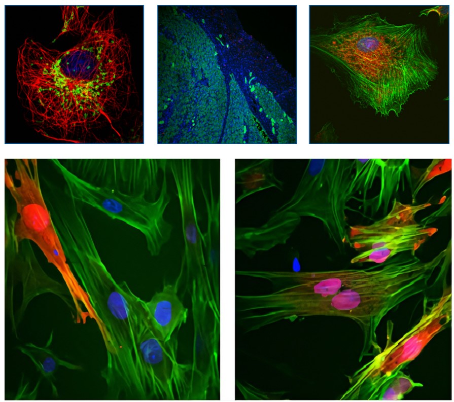
| Scanning confocal microscopy for the detection of cells in some plant and animal tissues |
Based on the diversity of its functions, LSCM is widely used in cell biology, physiology, neuroscience, genetics and other biological disciplines. In addition to conventional biology and medical applications, laser confocal microscopy is also gradually adopted in pharmacology,polymer science, paleoclimate stratigraphy,qualitative analysis of regenerated cellulose fibers and other emerging disciplines and it has been widely used.